.png)
Our Services
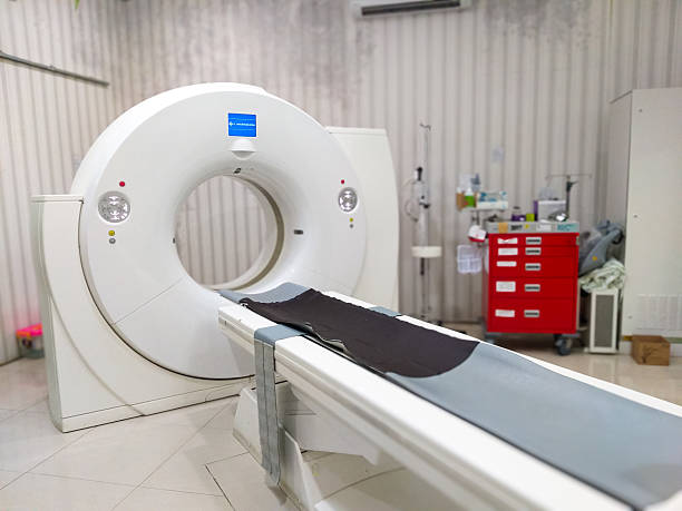
The nuclear medicine department at the center is equipped with advanced facilities and offers high-end services that support accurate and precise cancer diagnoses. Further, some of the highly experienced and the best nuclear medicine specialists in Region are available at the center and they are trained to deliver the highest quality nuclear medicine solutions.
Oncology (Cancer Imaging & Monitoring)
Early detection, accurate tumor localization, staging, and monitoring treatment response.
- Early detection and staging of cancers
- Monitoring treatment response and detecting recurrences
- Precise tumor localization with PET-CT
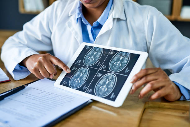
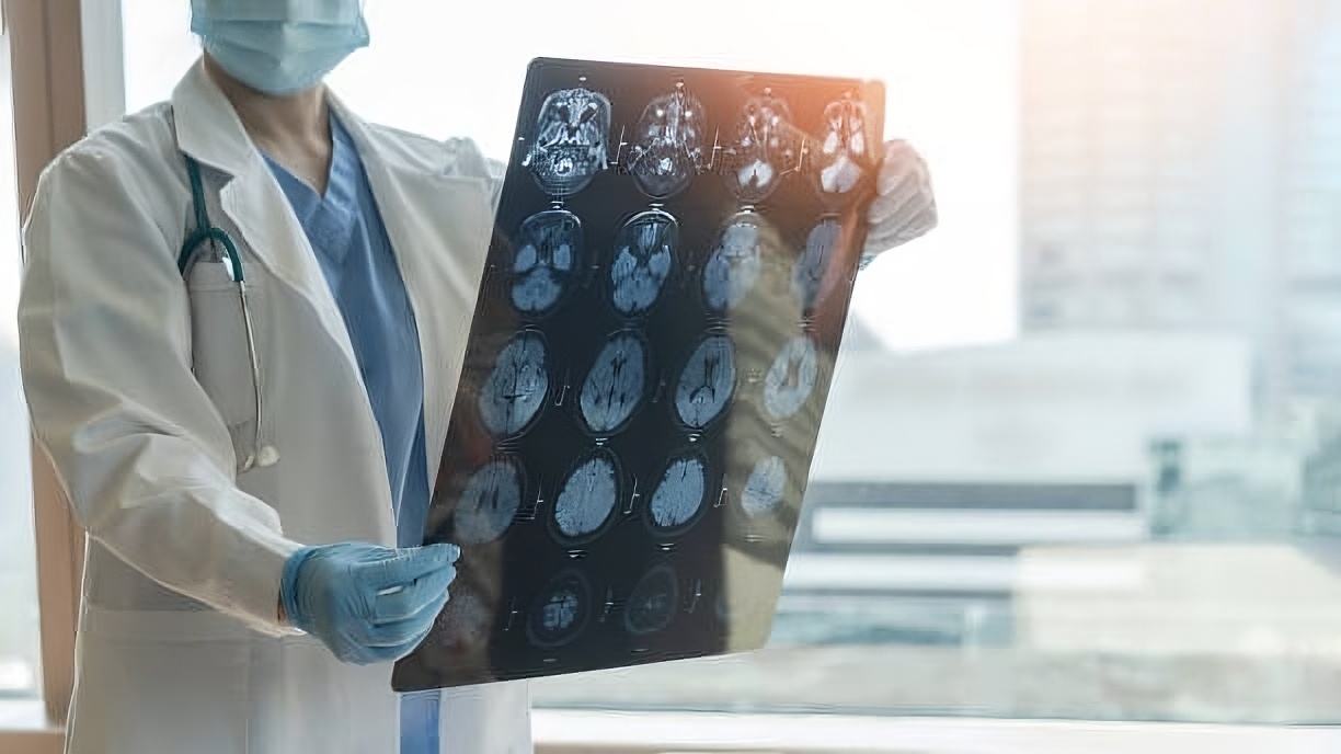
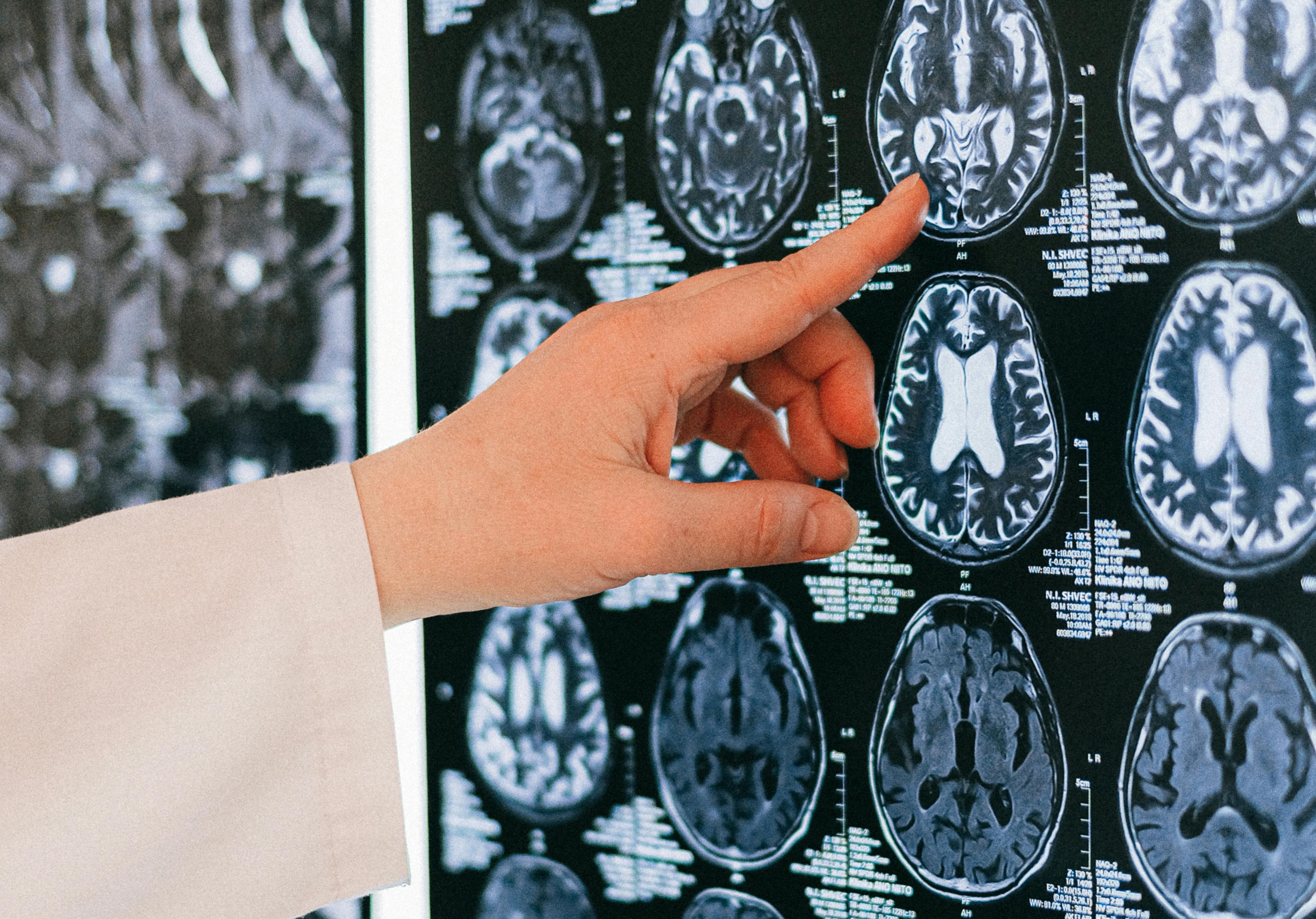
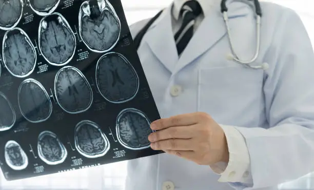
Neurology (Brain Disorders)
Assessment of Alzheimer’s, dementia, epilepsy, Parkinson’s, and brain tumors for precise diagnosis.
- Evaluation of Alzheimer’s, dementia, epilepsy, and Parkinson’s disease
- Assessment of brain tumors and neurological conditions
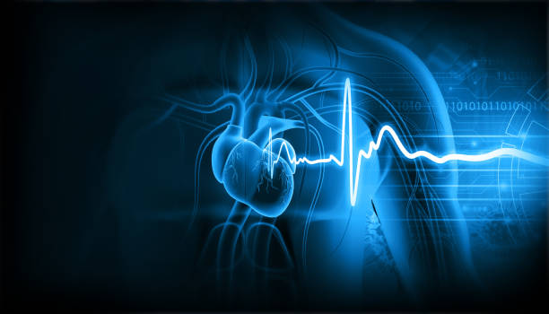
Cardiology (Heart Disease Imaging)
Evaluation of heart function, blood flow, and myocardial viability for effective treatment planning.
- Assessment of myocardial viability in coronary artery disease
- Evaluation of heart function and detection of abnormalities
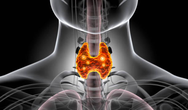
Thyroid and Endocrine PET Imaging
Detection of thyroid disorders, nodules, and endocrine tumors to guide targeted therapies.
- Assessment of thyroid disorders, nodules, and endocrine tumors
- Helps in planning targeted therapies
Infection and Inflammation Imaging
Identification of hidden infections or inflammation to support precise treatment.
- Detection of hidden infections or inflammatory conditions
- Supports targeted treatment planning
Advanced PET-CT Technology & Theranostics
Combining PET and CT for accurate diagnosis and personalized therapy planning.
- Combines PET (functional imaging) and CT (anatomical imaging) for highly accurate diagnosis
- Enables personalized therapy using theranostic approaches
PET – CT Scan With or Without MRI fusion
Center has the facility for performing PET-CT scans with or without MRI fusion.
- When performed with MRI fusion, the technique combines the benefits of all three imaging techniques.
- It enhances the diagnostic precision and helps in appropriately evaluating whether there is a need to change the treatment strategy.
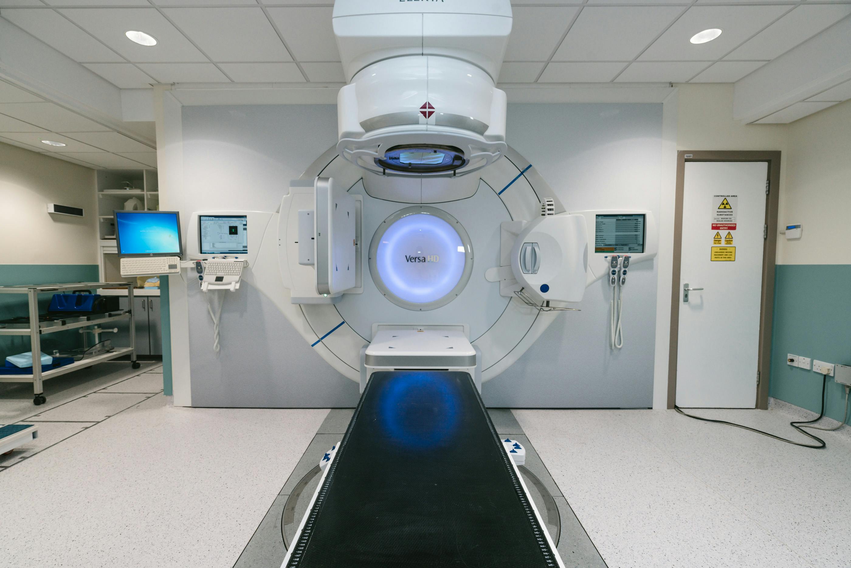
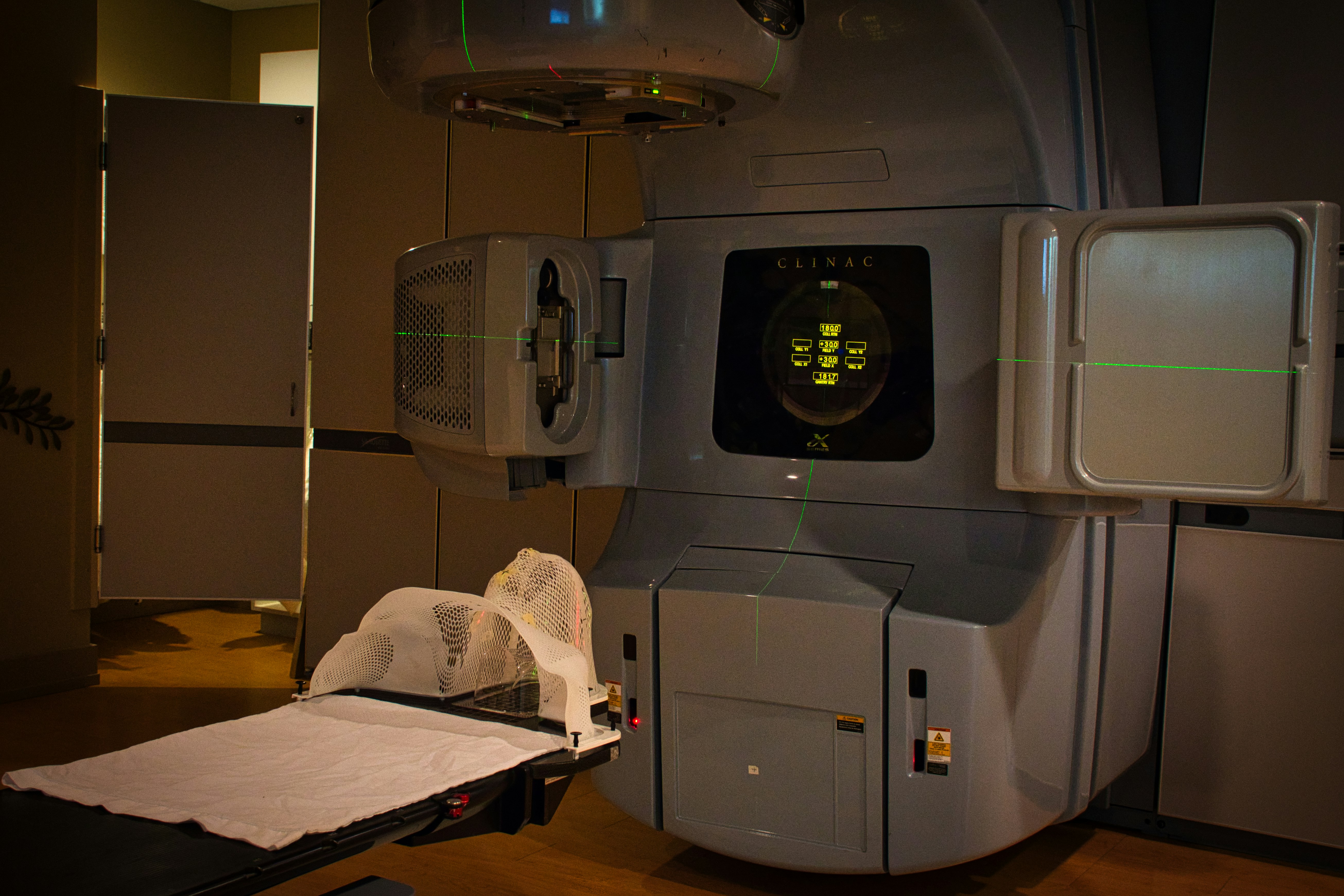
PET- CT-based Radiotherapy Planning
The center also has facilities for PET-CT-based radiotherapy planning.
- The surgeon plans radiotherapy based on the images obtained through the PET-CT scan.
- It improves the outcome and reduces healthy tissue damage.
PET- CT-based Treatment Response Evaluation
PET-CT results are used to determine the treatment response. .
- It helps the doctors in evaluating if they should be continuing the treatment or altering the treatment strategy.
PET- CT-based Biopsy Guidance
Compared to CT-guided Biopsy.
- PET-CT-guided biopsy is a highly effective and safe alternative for tumor diagnosis.
- Whole Body PET-CT: The facility of whole-body PET-CT is available at HCG Manavata Cancer Center; this allows doctors to screen and accurately diagnose cancer.
Cardiac PET-CT
MRI and echocardiography are two effective methods for diagnosing cardiac tumors.
- PET in combination with MRI or CT improves the overall diagnostic efficacy.
Radioactive Iodine Therapy
Facilities for radioactive iodine therapy, which is involved in treating several cancers, are also available Facilities.
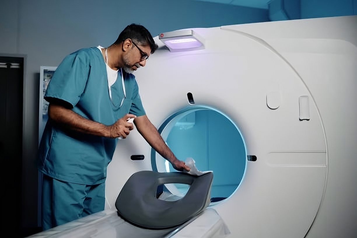
Magnetic Resonance Imaging
It is an advanced and rapid technology for diagnosing cancer.
- It involves using radio waves and magnets to develop clear images of the internal structures.
- The MRI does not use ionizing radiations.
SPECT
It is a nuclear medicine test
- that allows the doctor to evaluate the functioning of the internal structure.
- It also helps to determine the activity of a particular organ.
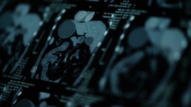
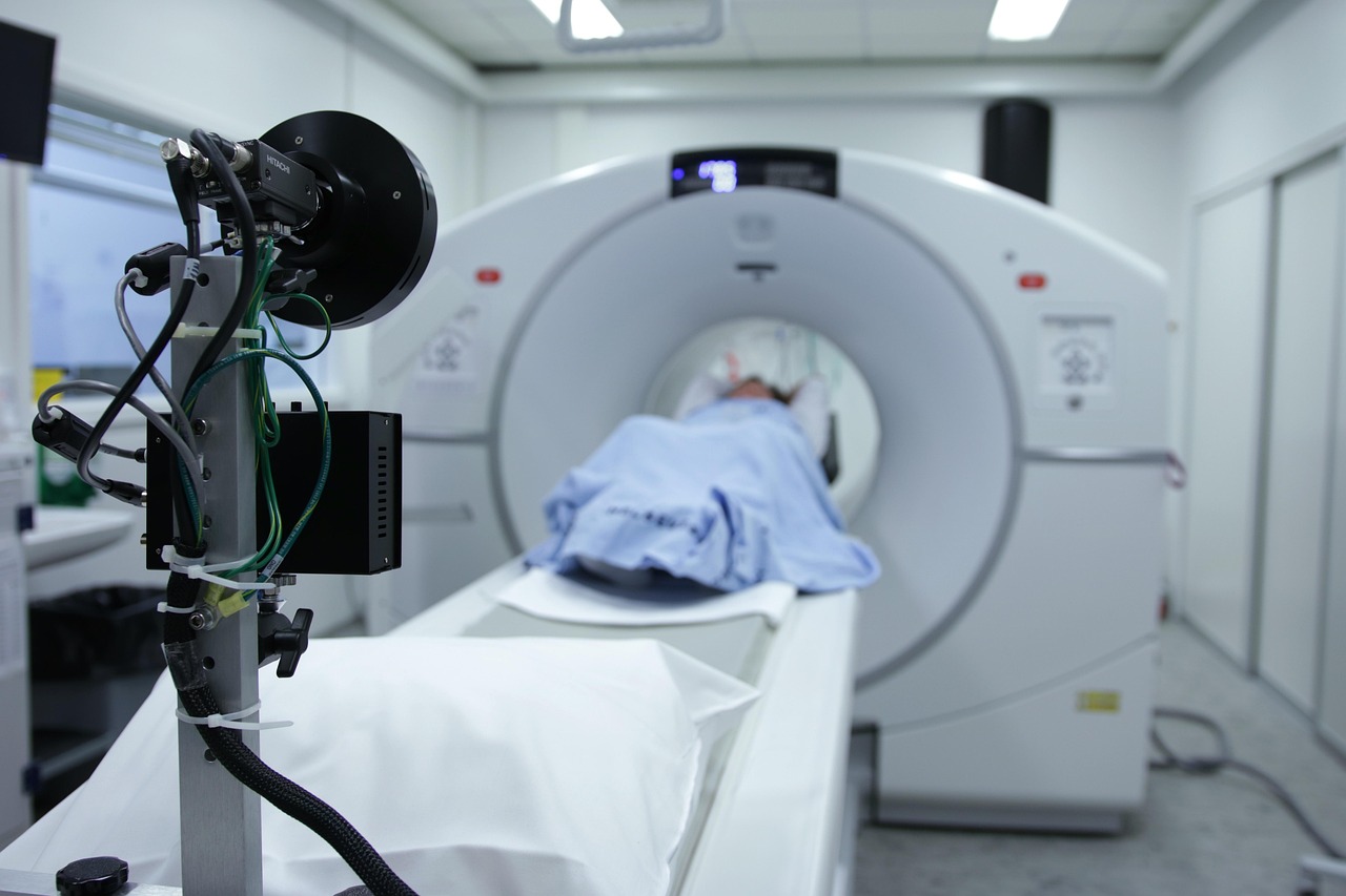
PET-CT
This scanning technology offers several advantages over either scanning technique alone.
- This technology provides the biochemical and structural details of the internal organs
- and this data helps technologists arrive at an accurate diagnosis for various health conditions.
SPECT (Gamma Camera)
What is SPECT?
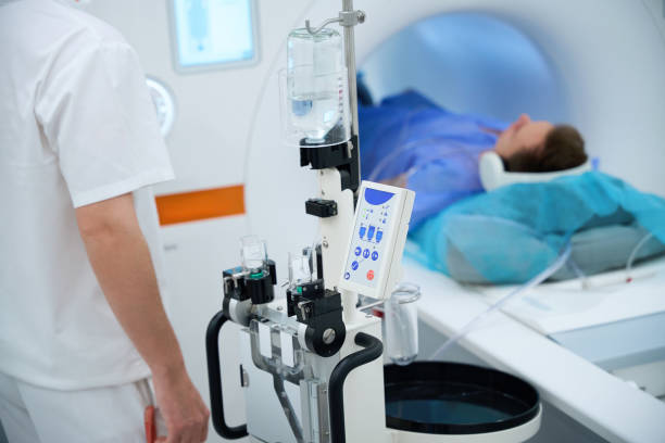
Single-photon emission computerized tomography (SPECT) scan is used to analyze the functions of the internal organs. A SPECT scan is a type of nuclear imaging test, which means it uses a radioactive substance and a special camera to create 3D pictures. While imaging tests such as X-rays can show what the structures inside your body look like, a SPECT scan produces images that show how your organs work. For instance, a SPECT scan can show how blood flows to your heart or what areas of your brain are more active or less active.
How does a SPET Scan Work?
A SPECT scan integrates two technologies, i.e computed tomography (CT) and a radioactive material (tracer) to view the body. Doctors can see how blood is pumped to tissues and organs because of the tracer. A radioactive substance is given through IV and the tracer dose is very small but the radioactive substance is absorbed more by the most active tissues. The SPECT machine camera detects the absorbed radioactive tracers in the body, it takes pictures and they are sent to a computer that creates a 3D image of the body or body part. Benefits of SPET CT at EMRALD PETCT CENTRE Patient experience: make the most of their time while lessening their dose Provide shorter, more tolerable exams for greater patient comfort, with Evolution technology. Streamlined exam setup leaves you with more time to interact with your patient while increasing throughput. Improves patient experience and enhances patient satisfaction.Our Dr Speaks persnoally with all patients and listen .
Why am I having a SPECT scan?
A SPECT Scan is used to diagnose or monitor disorders of brain, heart and bone. Is there any preparation on the day of the scan? What to bring and wear Bring all your previous reports especially Chest X-rays, PET Scans, CT & MRI Scan with Xerox of the CT/MRI written reports. Please bring your Doctor's note. Kindly remove metallic accessories and jewellery from your body.
Preparation
Arrive on time Please ensure your presence on scheduled appointment time. If you must cancel or reschedule, please do so at least 24 hours before your appointment. Your SPECT Scan After registering, you will go to a preparation area where a technologist will insert radioactive substance through an intravenous (IV) infusion into a vein in your arm. You will be asked to lie down for 20 minutes or more in the room, but you may feel cold sensation when the radioactive substance enters the body and it would be absorbed by the body. Sometimes it takes a few hours for it to be absorbed by the body In case of any extra or delayed scans are needed, the doctor/technologist will personally explain to you. After your scan Most of the radioactive tracer will be flushed out of your body through urine after few hours of scan. Yet some times one is instructed to drink more fluids, such as juice or water.
Radionuclide Scans
Our center provides a comprehensive range of radionuclide scans across multiple specialties for accurate diagnosis and treatment planning.
Gastroenterology
- Hepatobiliary scan
- Gastro-oesophageal reflux
- GI Bleed Scintigraphy
- Meckel’s diverticulum Scintigraphy
- Gastric emptying
- Blood pool study
Nephrourology
- Renogram
- Renal scan (DMSA-III)
- Vesicoureteric reflux
Pulmonary System
- Lung perfusion
Neurology
- Brain SPECT scan
Oncology
- Scintimammography
- Gallium scan
- Liver/Spleen SPECT scan
- MIBI scan
- DMSA–V Scan
Orthopaedics
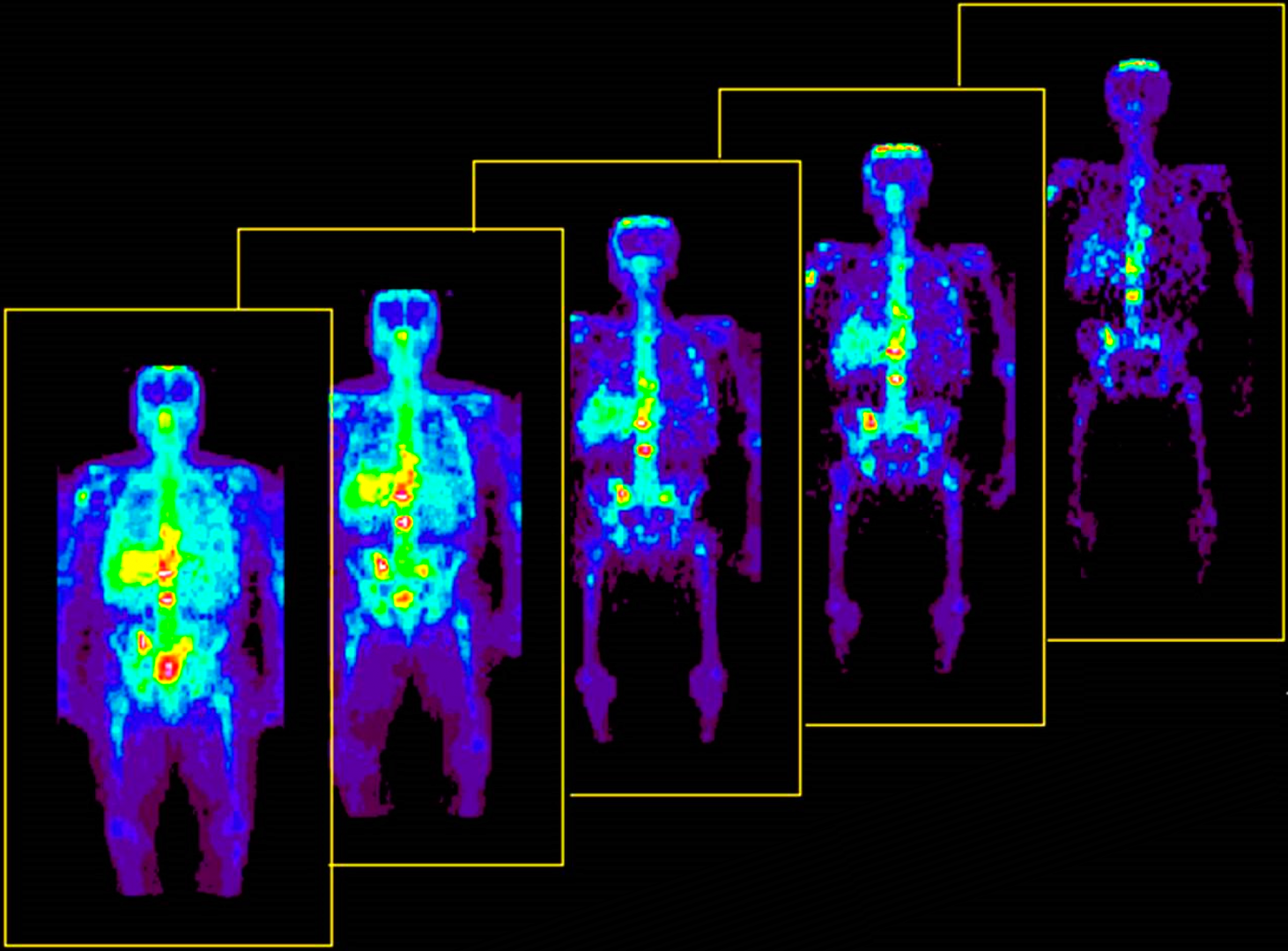
- Bone scan
- Bone graft viability
Cardiology
- Myocardial Perfusion Scintigraphy
- Myocardial viability
- LV ejection fraction (MUGA)
Endocrine
- Thyroid scan
- I-131 Whole body scan
- I-131 MIBG scan
- Parathyroid scintigraphy
Others
- Lymphoscintigraphy
- Gallium scan for infection
- Dacryoscintigraphy
- Salivary Scintigraphy
- Radionuclide Venogram
Radionuclide Therapy
- I-131 therapy for hyperthyroidism
- I-131 therapy for differentiated thyroid cancer
- Palliative treatment of painful bone metastasis
- P-32 therapy for Polycythemia Vera
.png)